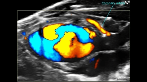
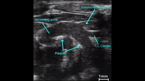
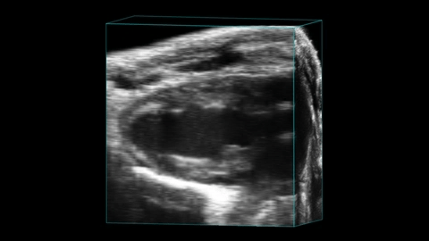
Now featuring the Vevo F2xc preclinical ultrasound system.
Training will be required before room and equipment privileges are granted. To begin, complete the "New User Registration" form.
|

Ranked13th nationally and 1stamong Big Ten by the Academy for Radiology & Biomedical Imaging Research
|
|
Overview of Services
|
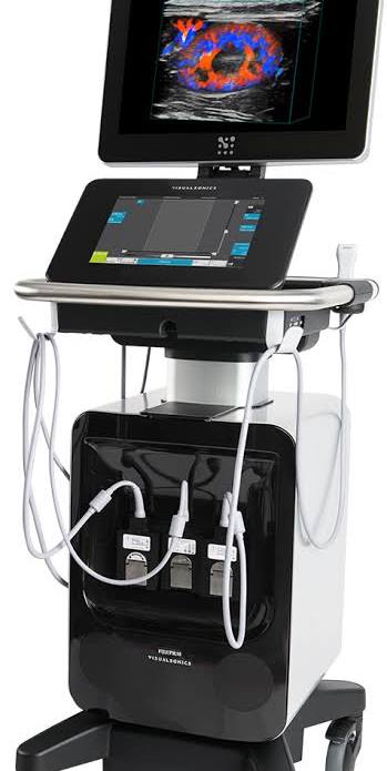
The Small Animal Ultrasound Core provides access to the F2-cx, a high-resolution digital imaging platform with linear technology and Color Doppler. The Vevo F2xc is ideally suited for cross-functional biological and physiological research. This flagship preclinical imaging platform allows for ultra-high to low frequency ultrasound imaging (71-1 MHz). Powered by next-generation electronics and system architecture, you can image a range of animal models at a low frequency for penetration, and ultra-high frequency for resolution—all in one platform.
The machine offers:
• 30micron resolution
• frame rates up to 10,000 frames per second
• Color and Power Doppler for blood flow quantification and anatomical identification.
• 4D Imaging, rendering, reconstruction & volume analysis
• Superb B-mode (2D) imaging with frame rates up to 10,000 fps with multiple focal zones for enhanced image uniformity
• M-mode assesses motion in cardiovascular applications
• Pulsed-wave Doppler (PW) for blood flow quantification
• Color Doppler shows flow direction, mean velocities & identifies small vessels not seen in B-mode
• Power Doppler for relative quantification of blood flow
• Simultaneous modes and steering for easier & faster studies
• MicroMarker™ Contrast Imaging – for relative perfusion & targeted molecular data
|
Probes Available:
UHF71x
UHF57x
UHF46x
UHF29x
UHF22x
P11-3
P5-1
Equipment fees for Fiscal Years 2027-2028
The Vevo2100 will be decommissioned before the end of 2026. The reduced rate for the 2100 is temporary. Information will be distributed closer to time.
- Vevo 2100 Unassisted Imaging-$33.86 F2xc Unassisted Imaging - $87.04
- Vevo 2100 Training and *Assisted Use-$85.94 F2xc Training and *Assisted Use - $139.11
- Vevo 2100 Data Analysis-$22.66 F2xc Analysis - $22.66
The FUJIFILM VisualSonics Vevo F2 ultrasound system was acquired by the Research Imaging Core within the Medical Imaging Research Institute in the Department of Radiology & Imaging Sciences and is now part of the new ultrasound research imaging core for the IU School of Medicine.
|
|
|
Leadership
| P. Melanie Pride |
R. Mark Payne |
| Ultrasound Core Manager |
Ultrasound Core Director |
| ppride@iu.edu |
|
Location and hours of operation
| Hours |
Location |
|
24/7
External users: 9am-5pm
|
| R2-177 |
| 950 W. Walnut St |
| Indianapolis, IN 46202 |
|
Links and Resources
- Visual Sonics F2xc Information Page
- F2xc Manual
- F2xc Brochure
- The FUJIFILM VisualSonics Learning Hub
- VevoStrain Application Software
- Self-Use Data Analysis Software VevoLab
- Transthoracic Echocardiography in Mice Video
- Publications Library | FUJIFILM VisualSonics
- Miss a workshop? Watch it Here On Demand | FUJIFILM VisualSonics
- Research Ultrasound Webinars | FUJIFILM VIsualSonics
- Animal Set-Up (litmos.com)
- Guide to Small Animal Intermediate Cardiac Imaging using Vevo Ultrasound Systems
 Welcome!
 ALERT
ALERT